
Cognard Type V Dural Arteriovenous Fistula: A Case Report
DOI:
https://doi.org/10.3941/jrcr.6185Abstract
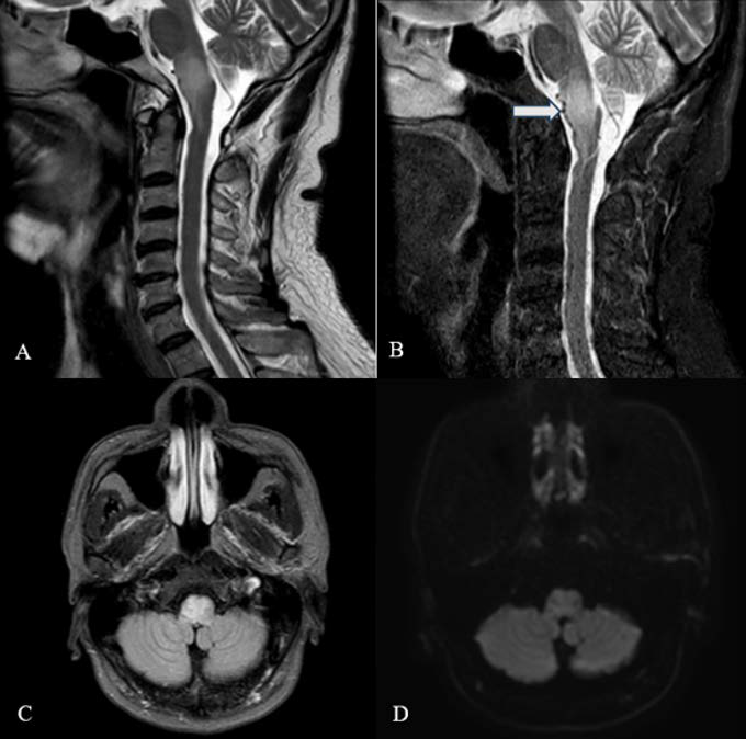
Dural arteriovenous fistula with perimedullary venous drainage (Cognard type V) is a rare intracranial vascular lesion that may cause progressive myelopathy due to venous hypertension and is frequently misdiagnosed because of nonspecific clinical and magnetic resonance imaging (MRI) findings. We report an adult patient presenting with progressive neurological deficits at the cervicomedullary junction. MRI showed diffuse T2-Weighted hyperintensity and swelling without diffusion restriction; subtle perimedullary flow voids were initially overlooked. The patient received corticosteroids for presumed myelitis, with subsequent deterioration. Digital subtraction angiography revealed a dural arteriovenous fistula supplied by intracranial dural branches with perimedullary venous drainage. Owing to unfavorable anatomy for endovascular therapy, microsurgical disconnection was performed. Therefore, early recognition and angiographic confirmation are essential to avoid delayed diagnosis and irreversible neurological injury.
Downloads
Published
Issue
Section
License
Copyright (c) 2026 Journal of Radiology Case Reports

This work is licensed under a Creative Commons Attribution-NonCommercial-NoDerivatives 4.0 International License.
The publisher holds the copyright to the published articles and contents. However, the articles in this journal are open-access articles distributed under the terms of the Creative Commons Attribution-NonCommercial-NoDerivs 4.0 License, which permits reproduction and distribution, provided the original work is properly cited. The publisher and author have the right to use the text, images and other multimedia contents from the submitted work for further usage in affiliated programs. Commercial use and derivative works are not permitted, unless explicitly allowed by the publisher.


