Retinoblastoma Metastatic to Bone: Case Series and Literature Review
DOI:
https://doi.org/10.3941/jrcr.6131Abstract
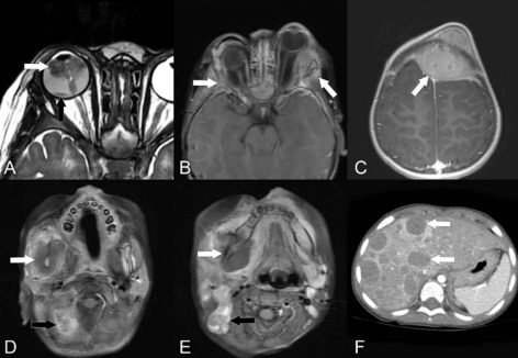
Retinoblastoma (RB) is a treatable tumor that arises in the retina, mainly affecting children under 2 years old. It results from a mutation on the long arm of chromosome 13, specifically of the RB1 gene (13q14). Patients with the hereditary form of RB have a higher incidence of metastases and secondary cancer development. These cancers, including metastasis, secondary malignant neoplasms, and radiation-associated sarcomas, are rare and typically occur in advanced stages. Although the overall prognosis for RB is generally favorable, it declines significantly when metastases develop. While rare, the most common sites of RB metastasis are the brain, osseous structures, cervical nodes, central nervous system, lungs, and liver. Here, we present the clinical and radiologic features of three patients with osseous metastasis from RB and review current literature, including the development of secondary malignant neoplasms, radiation-associated sarcomas, prognosis, and treatment options.
Downloads
Published
Issue
Section
License
Copyright (c) 2026 Journal of Radiology Case Reports

This work is licensed under a Creative Commons Attribution-NonCommercial-NoDerivatives 4.0 International License.
The publisher holds the copyright to the published articles and contents. However, the articles in this journal are open-access articles distributed under the terms of the Creative Commons Attribution-NonCommercial-NoDerivs 4.0 License, which permits reproduction and distribution, provided the original work is properly cited. The publisher and author have the right to use the text, images and other multimedia contents from the submitted work for further usage in affiliated programs. Commercial use and derivative works are not permitted, unless explicitly allowed by the publisher.