CT Characteristics and Pathological Correlation of Gastric Hamartomatous Inverted Polyp: A Case Report
DOI:
https://doi.org/10.3941/jrcr.6099Abstract
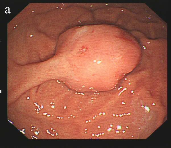
Gastric hamartomatous inverted polyp is a rare subtype of gastric polyp characterized pathologically by proliferation of submucosal gastric-type glands with cystic dilation and accompanying bundles of branching smooth muscle bundles, sometimes with infiltration of inflammatory cells. This report aims to improve understanding of the imaging features of gastric hamartomatous inverted polyp by correlating CT characteristics with pathological results.We present a case of a 51-year-old female patient. The patient underwent thorough endoscopy, endoscopic ultrasound, and CT examinations, and the postoperative pathology confirmed an inverted polyp in the stomach.The CT findings of gastric hamartomatous inverted polyp show a localized depression on top of the nodule, enhanced mucosal line that connects with the gastric mucosa, and small low-density cystic shadows inside the nodule, which closely correspond to the pathological changes.This case the complementary value of multiple imaging modalities in evaluating gastric lesions and improving diagnostic accuracy.
Downloads
Published
Issue
Section
License
Copyright (c) 2026 Journal of Radiology Case Reports

This work is licensed under a Creative Commons Attribution-NonCommercial-NoDerivatives 4.0 International License.
The publisher holds the copyright to the published articles and contents. However, the articles in this journal are open-access articles distributed under the terms of the Creative Commons Attribution-NonCommercial-NoDerivs 4.0 License, which permits reproduction and distribution, provided the original work is properly cited. The publisher and author have the right to use the text, images and other multimedia contents from the submitted work for further usage in affiliated programs. Commercial use and derivative works are not permitted, unless explicitly allowed by the publisher.