Multimodality Imaging of a Rare Case of Retroperitoneal Fetus in Fetu: A Radiologic Diagnostic Challenge
DOI:
https://doi.org/10.3941/jrcr.6035Abstract
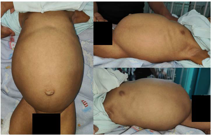
Introduction: Fetus in fetu is a rare congenital anomaly characterized by the presence of a malformed parasitic twin within its host, most commonly in the retroperitoneal space. Differentiation from mature teratomas is essential due to differences in malignant potential and management strategies. Case report: We report a 4-month-old male who presented with progressive abdominal distension, jaundice, and pale stools. Imaging studies revealed a large retroperitoneal mass with fetiform features. Imaging showed a retroperitoneal mass with organized skeletal structures compressing the biliary system. Surgical removal revealed a sac-like mass containing limbs and vertebral elements. Histopathology confirmed fetus in fetu without malignancy. Conclusion: Fetus in fetu should be considered in the differential diagnosis of complex retroperitoneal masses in infants. Multimodal imaging, especially the combination of ultrasonography, computed tomography, and magnetic resonance imaging, is invaluable for diagnosis, surgical planning, and distinguishing fetus in fetu from neoplastic entities.
Downloads
Published
Issue
Section
License
Copyright (c) 2026 Journal of Radiology Case Reports

This work is licensed under a Creative Commons Attribution-NonCommercial-NoDerivatives 4.0 International License.
The publisher holds the copyright to the published articles and contents. However, the articles in this journal are open-access articles distributed under the terms of the Creative Commons Attribution-NonCommercial-NoDerivs 4.0 License, which permits reproduction and distribution, provided the original work is properly cited. The publisher and author have the right to use the text, images and other multimedia contents from the submitted work for further usage in affiliated programs. Commercial use and derivative works are not permitted, unless explicitly allowed by the publisher.