
Occult Breast Cancer Presenting as Isolated Axillary Lymphadenopathy: A Case Report with MRI–Pathology Correlation
DOI:
https://doi.org/10.3941/jrcr.6021Abstract
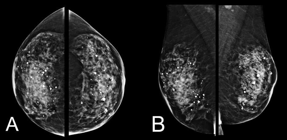
Occult breast cancer is a rare presentation of invasive ductal carcinoma in which axillary lymph node metastasis is identified without a detectable breast lesion on mammography or ultrasound. We report a case of a 57-year-old woman recalled from screening mammography for bilateral breast asymmetries that resolved on diagnostic compression views. Targeted ultrasound of the axillary revealed a solitary lymph node with progressive cortical thickening. Core needle biopsy demonstrated metastatic carcinoma, consistent with mammary origin. Breast MRI subsequently localized enhancing foci, confirmed as invasive ductal carcinoma. This case highlights the diagnostic value of contrast-enhanced breast MRI in identifying otherwise occult primary breast tumors and correlation of imaging and histopathologic findings in establishing a definitive diagnosis in clinical occult presentations.
Downloads
Published
Issue
Section
License
Copyright (c) 2026 Journal of Radiology Case Reports

This work is licensed under a Creative Commons Attribution-NonCommercial-NoDerivatives 4.0 International License.
The publisher holds the copyright to the published articles and contents. However, the articles in this journal are open-access articles distributed under the terms of the Creative Commons Attribution-NonCommercial-NoDerivs 4.0 License, which permits reproduction and distribution, provided the original work is properly cited. The publisher and author have the right to use the text, images and other multimedia contents from the submitted work for further usage in affiliated programs. Commercial use and derivative works are not permitted, unless explicitly allowed by the publisher.


