
A Case Report of Endovascular Embolization for Giant Congenital Hemangioma with Arteriovenous Fistula in A Neonate
DOI:
https://doi.org/10.3941/jrcr.6017Abstract
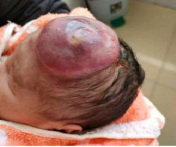
Background: Congenital hemangioma (CH) is a vascular tumor that develops during fetal life. Although most CHs are benign, those associated with high-flow arteriovenous fistulas (AVFs) can lead to life-threatening complications such as high-output cardiac failure and severe pulmonary hypertension. Case Presentation: We report a male preterm infant (birth weight: 2.8 kg) with a giant CH and AVF on the left forehead, confirmed by transcranial Doppler and magnetic resonance angiography (MRA). The patient rapidly developed high-output cardiac failure and respiratory failure. Despite maximal medical therapy, his condition deteriorated. A multidisciplinary team deemed surgical resection prohibitively high-risk, and transcatheter arterial embolization was selected as the primary intervention. Intervention and Outcome: On day 11 of life, successful embolization was performed via a right femoral approach under general anesthesia. Despite a femoral artery diameter <1 mm, vascular access was achieved using a pediatric needle and Seldinger technique. Angiography confirmed a high-flow AV shunt, which was embolized using a combination of detachable and free coils. Post-procedurally, pulmonary artery pressure decreased from 77 mmHg to 42 mmHg, and the ductus arteriosus shunt converted from right-to-left to bidirectional. The patient was weaned from mechanical ventilation on day 8 post-procedure. Four months later, radical resection confirmed CH with AV malformation. At one-year follow-up, there was no neurological deficit or recurrence. Conclusion: Transcatheter embolization is a life-saving treatment for neonates with CH and high-flow AVF when surgery is not feasible. This case highlights the feasibility of complex endovascular interventions in extremely low-weight infants and underscores the value of multidisciplinary management.
Downloads
Published
Issue
Section
License
Copyright (c) 2026 Journal of Radiology Case Reports

This work is licensed under a Creative Commons Attribution-NonCommercial-NoDerivatives 4.0 International License.
The publisher holds the copyright to the published articles and contents. However, the articles in this journal are open-access articles distributed under the terms of the Creative Commons Attribution-NonCommercial-NoDerivs 4.0 License, which permits reproduction and distribution, provided the original work is properly cited. The publisher and author have the right to use the text, images and other multimedia contents from the submitted work for further usage in affiliated programs. Commercial use and derivative works are not permitted, unless explicitly allowed by the publisher.


