
Leukoencephalopathy From SDHAF1-Related Mitochondrial Deficiency
DOI:
https://doi.org/10.3941/jrcr.5293Abstract
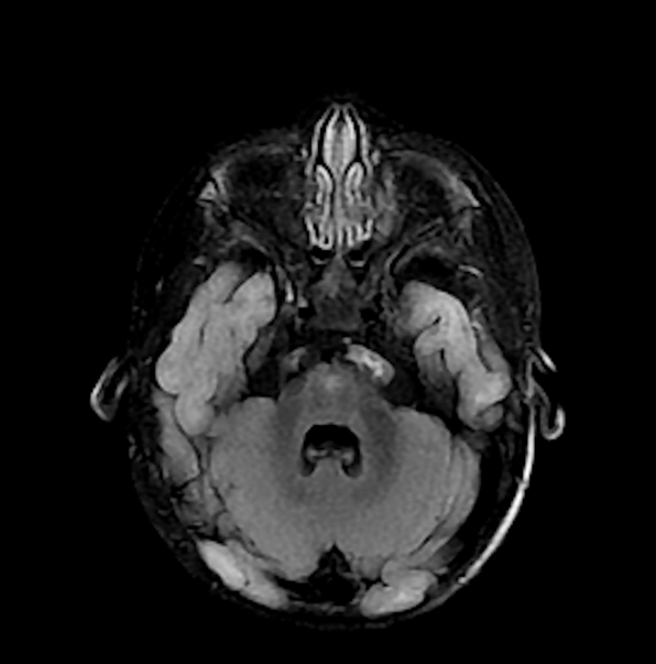
We present exceedingly rare cases of leukoencephalopathy due to SDHAF1-related mitochondrial complex II deficiency in identical twins. These findings contribute to the scarcity of reports present in both the radiology literature and the medical literature describing imaging characteristics associated with this condition. Like other mitochondrial disorders affecting the CNS, this disease typically presents in infants or young children with symptoms of weakness, hypotonia, and developmental regression. However, the imaging findings of SDHAF1-related mitochondrial complex deficiency in this pair of patients are unique and include symmetric white matter diffusion restriction most pronounced in the genu and splenium of the corpus callosum with sparing of the body. We propose that the specific imaging findings described in this report may be considered pathognomonic and may therefore be added to the list of previous neuroimaging findings associated with this entity. Recognition of this distinctive pattern by radiologists would allow for a prospective diagnosis on imaging that can then be confirmed with genomic testing and magnetic resonance spectroscopy analysis.
Downloads
Published
Issue
Section
License
Copyright (c) 2026 Journal of Radiology Case Reports

This work is licensed under a Creative Commons Attribution-NonCommercial-NoDerivatives 4.0 International License.
The publisher holds the copyright to the published articles and contents. However, the articles in this journal are open-access articles distributed under the terms of the Creative Commons Attribution-NonCommercial-NoDerivs 4.0 License, which permits reproduction and distribution, provided the original work is properly cited. The publisher and author have the right to use the text, images and other multimedia contents from the submitted work for further usage in affiliated programs. Commercial use and derivative works are not permitted, unless explicitly allowed by the publisher.


