
Erdheim-Chester disease on 18F-FDG PET/CT:A case report
DOI:
https://doi.org/10.3941/jrcr.5771Abstract
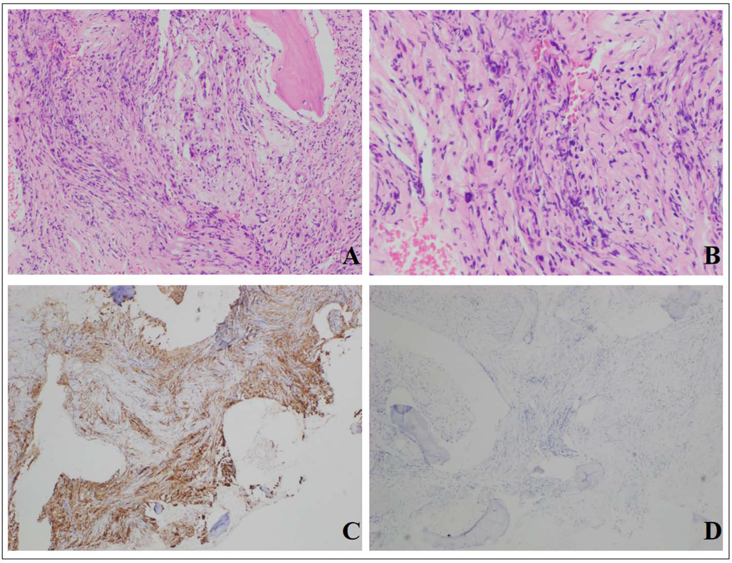
Erdheim-Chester disease (ECD) is a rare form of systemic non-Langerhans cell histiocytosis with characteristic bone involvement. Extraskeletal involvement sites were found in central nervous system (CNS), cardiovascular system, lungs, kidneys, and so on. The most common site of CNS involvement is the hypothalamic-pituitary axis, but here we report a rare case of ECD with the frontal lobe involvement.
Downloads
Published
Issue
Section
License
Copyright (c) 2025 Journal of Radiology Case Reports

This work is licensed under a Creative Commons Attribution-NonCommercial-NoDerivatives 4.0 International License.
The publisher holds the copyright to the published articles and contents. However, the articles in this journal are open-access articles distributed under the terms of the Creative Commons Attribution-NonCommercial-NoDerivs 4.0 License, which permits reproduction and distribution, provided the original work is properly cited. The publisher and author have the right to use the text, images and other multimedia contents from the submitted work for further usage in affiliated programs. Commercial use and derivative works are not permitted, unless explicitly allowed by the publisher.


