
Rare Combination of Frontonasal and Bilateral Naso-orbital Encephaloceles
DOI:
https://doi.org/10.3941/jrcr.v5i11.715Keywords:
Encephalocele, Neuroradiology, Pediatric radiology, frontonasal, naso-orbitalAbstract
Encephaloceles, while a common entity affecting 1:4000 live births, typically occur in the occipital region. Encephaloceles involving the frontal region comprise only 15% of all cases. Naso-orbital encephaloceles are rarely seen. Our case profiles a child born at term with an atrial septal defect (ASD), micrognathia, cleft lip, and frontonasal as well as bilateral naso-orbital encephaloceles. At birth the encephaloceles were undetected. During the cleft palate pre-operative preparation, the bilateral naso-orbital encephaloceles were diagnosed as dacrocystoceles for which the child underwent surgical repair. Misdiagnosis and loss to follow up lead to delayed surgical treatment until the child was almost two years of age; the right eye was near complete closure due to the increasing size of the encephalocele. This case highlights the importance of meticulous radiologic interpretation of midline nasal masses, as a correct diagnosis impacts clinical management and directs surgical repair.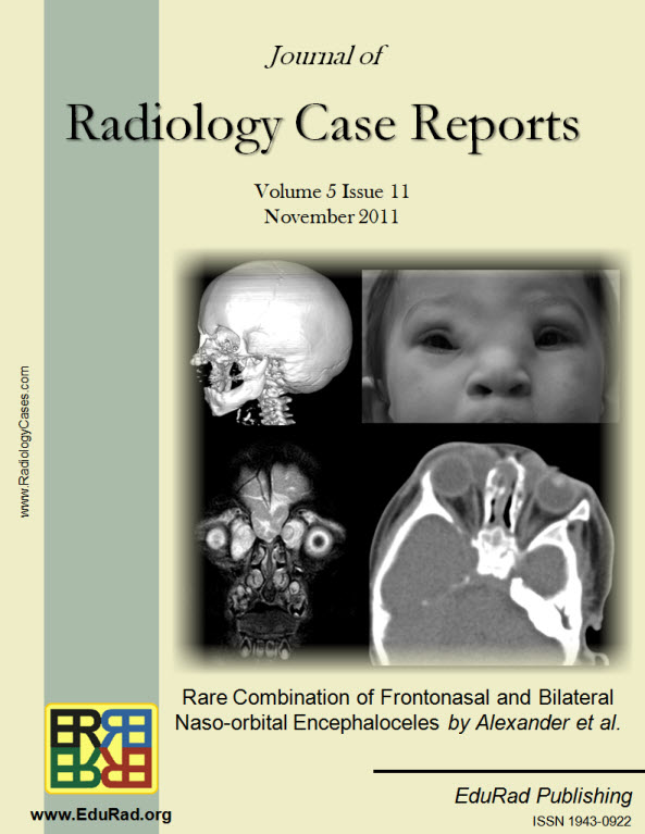
Downloads
Published
2011-11-12
Issue
Section
Pediatric Radiology
License
The publisher holds the copyright to the published articles and contents. However, the articles in this journal are open-access articles distributed under the terms of the Creative Commons Attribution-NonCommercial-NoDerivs 4.0 License, which permits reproduction and distribution, provided the original work is properly cited. The publisher and author have the right to use the text, images and other multimedia contents from the submitted work for further usage in affiliated programs. Commercial use and derivative works are not permitted, unless explicitly allowed by the publisher.


