Splenunculus Mimicking Neuroendocrine Tumour on Ga-68 DOTATATE Positron Emission Topography: The Diagnostic Value of Heat-Damaged Red Blood Cell Scintigraphy
DOI:
https://doi.org/10.3941/jrcr.6270Abstract
Background: Accessory splenic tissue can mimic pancreatic or peritoneal neoplasms on both cross-sectional and functional imaging, complicating diagnosis and surveillance. Technetium-99m heat-damaged red blood cell (HDRBC) scintigraphy is a highly specific functional imaging modality for identifying ectopic splenic tissue.
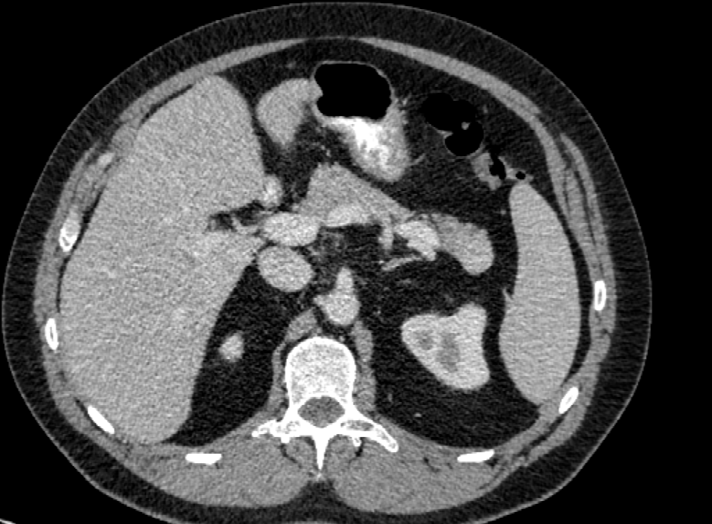
Case Summary: A 54-year-old man presented with non-specific gastrointestinal symptoms and was found to have a pancreatic tail lesion on contrast-enhanced CT of the abdomen and pelvis. Subsequent pancreatic MRI demonstrated signal characteristics similar to the adjacent spleen, raising the possibility of an intrapancreatic accessory splenule. Functional imaging with Gallium-68 (Ga-68) DOTATATE PET/CT demonstrated avid uptake in the pancreatic tail lesion, as well as additional DOTATATE-avid foci in the left upper quadrant peritoneum and terminal ileum, raising concern for multifocal neuroendocrine tumour. Surgical resection of the pancreatic tail lesion was performed, with histopathology confirming benign accessory splenic tissue. Surveillance imaging demonstrated persistent DOTATATE uptake in the terminal ileum and peritoneal nodule. Tc-99m heat-damaged red blood cell (HDRBC) scintigraphy confirmed the peritoneal lesion as functioning accessory splenic tissue. The patient underwent right hemicolectomy, with histopathology confirming a distal ileal neuroendocrine tumour. Follow-up Ga-68 DOTATATE PET/CT demonstrated no evidence of residual or recurrent malignancy, with stable uptake confined to known accessory splenic tissue.
Conclusion: This case illustrates the diagnostic challenge posed by accessory splenunculi on somatostatin receptor imaging and highlights the value of heat-damaged red blood cell scintigraphy in diagnosing accessory splenic tissue, which can often mimic neuroendocrine tumours on Gallium-68 DOTATATE PET/CT. It should be considered in the diagnostic algorithm for DOTATATE-avid lesions, to prevent misinterpretation and unnecessary intervention.
Downloads
Published
Issue
Section
License
Copyright (c) 2026 Journal of Radiology Case Reports

This work is licensed under a Creative Commons Attribution-NonCommercial-NoDerivatives 4.0 International License.
The publisher holds the copyright to the published articles and contents. However, the articles in this journal are open-access articles distributed under the terms of the Creative Commons Attribution-NonCommercial-NoDerivs 4.0 License, which permits reproduction and distribution, provided the original work is properly cited. The publisher and author have the right to use the text, images and other multimedia contents from the submitted work for further usage in affiliated programs. Commercial use and derivative works are not permitted, unless explicitly allowed by the publisher.