A Very Rare Cause of VP Shunt Dysfunction Following Contrast Injection into the Peritoneal Catheter during an Intra- Abdominal Procedure: A Case Report
DOI:
https://doi.org/10.3941/jrcr.6164Abstract
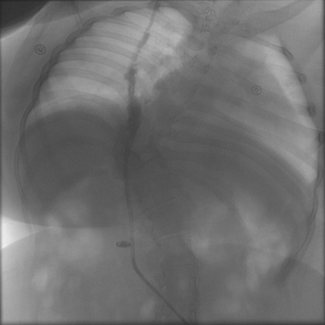
Background: Ventriculoperitoneal (VP) shunts are widely used for lifelong management of hydrocephalus. While early and late causes of VP shunt dysfunction are well described, very late mechanical failures due to unusual mechanisms remain rare. Case Presentation: We report a 24-year-old female with congenital hydrocephalus and myelomeningocele who underwent VP shunt insertion at birth. Following multiple complex urologic procedures, she developed VP shunt dysfunction secondary to inadvertent contrast injection into the distal peritoneal catheter, resulting in catheter rupture and subcutaneous leakage. She was successfully managed with shunt externalization, targeted antimicrobial therapy, and delayed VP shunt reinsertion. Conclusion: This case highlights a rare and preventable mechanism of very late VP shunt dysfunction related to radiologic contrast administration. Awareness of VP shunt anatomy and multidisciplinary coordination are essential when managing shunt-dependent patients undergoing complex urologic interventions.
Downloads
Published
Issue
Section
License
Copyright (c) 2026 Journal of Radiology Case Reports

This work is licensed under a Creative Commons Attribution-NonCommercial-NoDerivatives 4.0 International License.
The publisher holds the copyright to the published articles and contents. However, the articles in this journal are open-access articles distributed under the terms of the Creative Commons Attribution-NonCommercial-NoDerivs 4.0 License, which permits reproduction and distribution, provided the original work is properly cited. The publisher and author have the right to use the text, images and other multimedia contents from the submitted work for further usage in affiliated programs. Commercial use and derivative works are not permitted, unless explicitly allowed by the publisher.