A Rare Case of Kimura’s Disease Involving the Parotid Gland: CT and Diffusion MRI
DOI:
https://doi.org/10.3941/jrcr.6153Abstract
Kimura’s disease is a rare chronic inflammatory disorder that may involve the parotid gland and closely mimic neoplastic or
lymphoproliferative conditions on imaging. We describe the case of a 47-year-old man with parotid gland involvement, evaluated using
computed tomography and multiparametric magnetic resonance imaging, with emphasis on diffusion-weighted imaging and quantitative
apparent diffusion coefficient analysis. Computed tomography demonstrated a poorly defined soft-tissue mass within the parotid region,
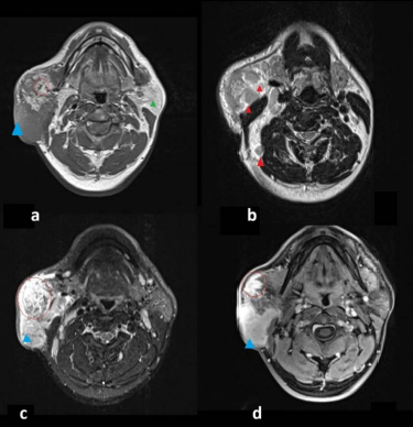
associated with in traparotid and cervical lymphadenopathy and heterogeneous contrast enhancement. Magnetic resonance imaging
revealed heterogeneous signal intensity, including relatively low T2 signal, and early and progressive, relatively homogeneous enhancement
suggestive of an inflammatory process. Quantitative diffusion analysis showed intermediate apparent diffusion coefficient values, consistent
with inflammatory tissue rather than highly cellular malignant lesions. Histogram analysis showed a moderately asymmetric distribution,
supporting a heterogeneous but non-neoplastic microstructural composition. These findings differed from the typical diffusion patterns
of lymphoma and pleomorphic adenoma, with only partial overlap with ductal carcinoma. This case highlights the complementary role
of computed tomography and multiparametric magnetic resonance imaging, particularly diffusion-weighted imaging with quantitative
apparent diffusion coefficient assessment, in the noninvasive differential diagnosis of rare inflammatory diseases affecting the parotid gland.
Downloads
Published
Issue
Section
License
Copyright (c) 2026 Journal of Radiology Case Reports

This work is licensed under a Creative Commons Attribution-NonCommercial-NoDerivatives 4.0 International License.
The publisher holds the copyright to the published articles and contents. However, the articles in this journal are open-access articles distributed under the terms of the Creative Commons Attribution-NonCommercial-NoDerivs 4.0 License, which permits reproduction and distribution, provided the original work is properly cited. The publisher and author have the right to use the text, images and other multimedia contents from the submitted work for further usage in affiliated programs. Commercial use and derivative works are not permitted, unless explicitly allowed by the publisher.