
A Pitfall of Fluorodeoxyglucose Positron Emission Tomography/Computed Tomography: Fecal Peritonitis Mimicking Peritoneal Carcinomatosis
DOI:
https://doi.org/10.3941/jrcr.6001Abstract
Background: Postoperative inflammatory reactions, such as foreign-body responses, can mimic peritoneal carcinomatosis on fluorodeoxyglucose positron emission tomography, potentially leading to misdiagnosis.
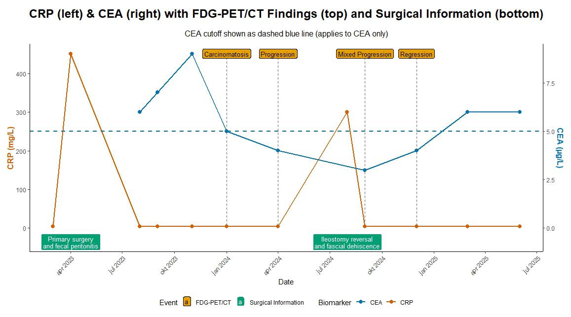
Case presentation: A 65-year-old woman with colon adenocarcinoma developed fecal peritonitis after surgery. Rising carcinoembryonic antigen and imaging findings suggested peritoneal carcinomatosis. Repeated biopsies revealed inflammatory tissue with foreign material consistent with prior peritonitis. Follow-up imaging and biomarker normalization confirmed a benign etiology despite persistent uptake.
Conclusion: Interpretation of postoperative fluorodeoxyglucose positron emission tomography requires integration of imaging, histology, and clinical context to avoid unnecessary treatment.
Downloads
Published
Issue
Section
License
Copyright (c) 2026 Journal of Radiology Case Reports

This work is licensed under a Creative Commons Attribution-NonCommercial-NoDerivatives 4.0 International License.
The publisher holds the copyright to the published articles and contents. However, the articles in this journal are open-access articles distributed under the terms of the Creative Commons Attribution-NonCommercial-NoDerivs 4.0 License, which permits reproduction and distribution, provided the original work is properly cited. The publisher and author have the right to use the text, images and other multimedia contents from the submitted work for further usage in affiliated programs. Commercial use and derivative works are not permitted, unless explicitly allowed by the publisher.


