
Scapulothoracic Bursitis in a Patient with Rheumatoid Arthritis: Multimodality Imaging and Spontaneous Resolution
DOI:
https://doi.org/10.3941/jrcr.5980Abstract
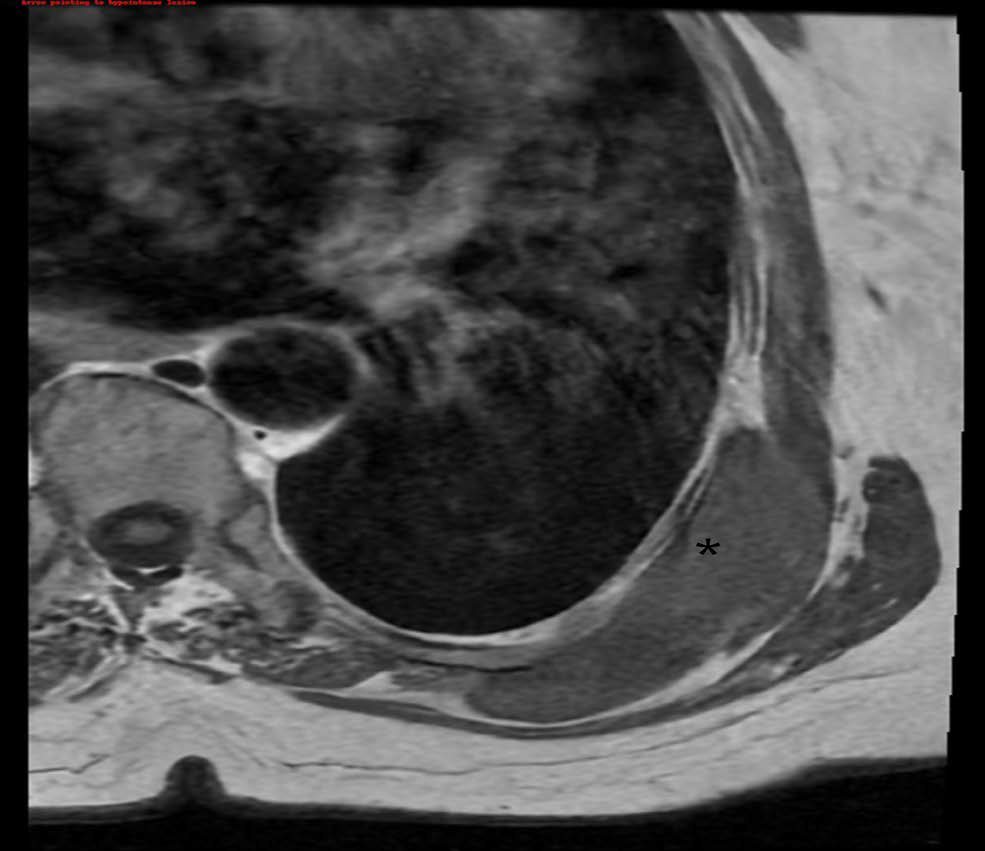
Scapulothoracic bursitis is an uncommon condition characterized by inflammation of the bursa located between the scapula and the thoracic wall. This disorder is frequently associated with repetitive shoulder movements or systemic inflammatory diseases. This report describes a 56-year-old woman with rheumatoid arthritis who presented with painless swelling in the left upper back. Imaging studies identified a well-defined, fluid-filled lesion beneath the latissimus dorsi and serratus anterior muscles. Ultrasound demonstrated an anechoic lesion without vascularity, and magnetic resonance imaging (MRI) confirmed a T1 hypointense and T2 hyperintense lesion with fine septations. The lesion resolved spontaneously without surgical intervention, possibly facilitated by ongoing anti-inflammatory therapy. This case emphasizes the need to consider scapulothoracic bursitis in the differential diagnosis of posterior chest wall masses and demonstrates the critical role of imaging in preventing unnecessary invasive procedures.
Downloads
Published
Issue
Section
License
Copyright (c) 2026 Journal of Radiology Case Reports

This work is licensed under a Creative Commons Attribution-NonCommercial-NoDerivatives 4.0 International License.
The publisher holds the copyright to the published articles and contents. However, the articles in this journal are open-access articles distributed under the terms of the Creative Commons Attribution-NonCommercial-NoDerivs 4.0 License, which permits reproduction and distribution, provided the original work is properly cited. The publisher and author have the right to use the text, images and other multimedia contents from the submitted work for further usage in affiliated programs. Commercial use and derivative works are not permitted, unless explicitly allowed by the publisher.


