
Imaging Characteristics of Angiomatoid Fibrous Histiocytoma: A Case Report and the Diagnostic Challenges in Differentiating Fluid–Fluid Level Soft Tissue Lesions
DOI:
https://doi.org/10.3941/jrcr.5959Abstract
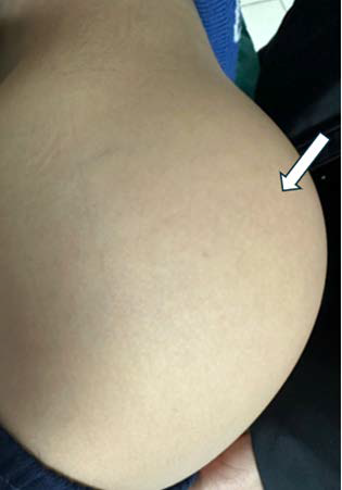
Angiomatoid fibrous histiocytoma (AFH) is a rare mesenchymal tumor. Due to its non-specific clinical and radiologic features, AFH is often misdiagnosed as other soft tissue lesions. We report the case of a 10-year-old boy who presented with a painless mass in the gluteal region. Initial ultrasound and Magnetic Resonance Imaging (MRI) suggested a hemorrhagic lesion with fluid–fluid levels. Despite two inconclusive percutaneous biopsies, the patient underwent wide local excision, and histopathological examination confirmed the diagnosis of AFH. This case highlights the diagnostic challenges of AFH and underscores the importance of recognizing its imaging characteristics—particularly in distinguishing it from other soft tissue lesions that also exhibit fluid–fluid levels on MRI—in the appropriate clinical context.
Downloads
Published
Issue
Section
License
Copyright (c) 2025 Journal of Radiology Case Reports

This work is licensed under a Creative Commons Attribution-NonCommercial-NoDerivatives 4.0 International License.
The publisher holds the copyright to the published articles and contents. However, the articles in this journal are open-access articles distributed under the terms of the Creative Commons Attribution-NonCommercial-NoDerivs 4.0 License, which permits reproduction and distribution, provided the original work is properly cited. The publisher and author have the right to use the text, images and other multimedia contents from the submitted work for further usage in affiliated programs. Commercial use and derivative works are not permitted, unless explicitly allowed by the publisher.


