
Atypical IVA Hepatic Duct Cyst Mimicking Biliary Atresia: The Critical Role of Multimodal Imaging and its Diagnostic Pitfalls
DOI:
https://doi.org/10.3941/jrcr.5924Abstract
Biliary tree cysts are rare congenital anomalies, characterized by cystic dilatation of intrahepatic and/or extrahepatic bile ducts. Their incidence is approximately 1 case in 100,000-150,000 live births in western populations, notably more frequent in east Asia. Although most cases are diagnosed in the early childhood, up to 30% of the cases present in the neonatal period, often manifesting with prolonged jaundice as a nonspecific clinical feature. Their prompt differentiation from cystic biliary atresia is critical, due to its different management and prognosis.
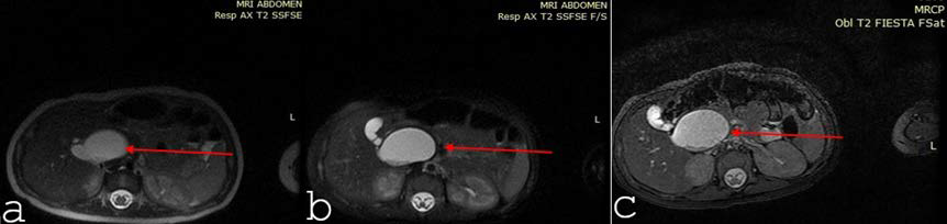
We present the case of a 2-day old male neonate with conjugated hyperbilirubinemia, jaundice and abdominal distension. Ultrasonography revealed a cystic lesion distinct from the gallbladder, and further abdominal MRI and MRCP confirmed an atypical type IVA right hepatic duct cyst, without extrahepatic duct involvement. The case posed a diagnostic challenge due to overlapping clinical features with cystic biliary atresia. Early recognition of the accurate diagnosis, allowed for timely surgical planning, preventing complications as cholangitis, pancreatitis, and potential long-term malignant transformation.
This case accentuates the importance of maintaining broad differential diagnosis in neonatal cholestasis, and demonstrates how integrated imaging can accurately distinguish hepatic duct cysts from cystic biliary atresia. Moreover, early surgical referral is essential to mitigate long-term hepatobiliary morbidity.
Downloads
Published
Issue
Section
License
Copyright (c) 2026 Journal of Radiology Case Reports

This work is licensed under a Creative Commons Attribution-NonCommercial-NoDerivatives 4.0 International License.
The publisher holds the copyright to the published articles and contents. However, the articles in this journal are open-access articles distributed under the terms of the Creative Commons Attribution-NonCommercial-NoDerivs 4.0 License, which permits reproduction and distribution, provided the original work is properly cited. The publisher and author have the right to use the text, images and other multimedia contents from the submitted work for further usage in affiliated programs. Commercial use and derivative works are not permitted, unless explicitly allowed by the publisher.


