
An Eosinophilic Pneumonia Mimicking Lung Cancer on Multiple Imaging Modalities Monitored By CT
DOI:
https://doi.org/10.3941/jrcr.5896Abstract
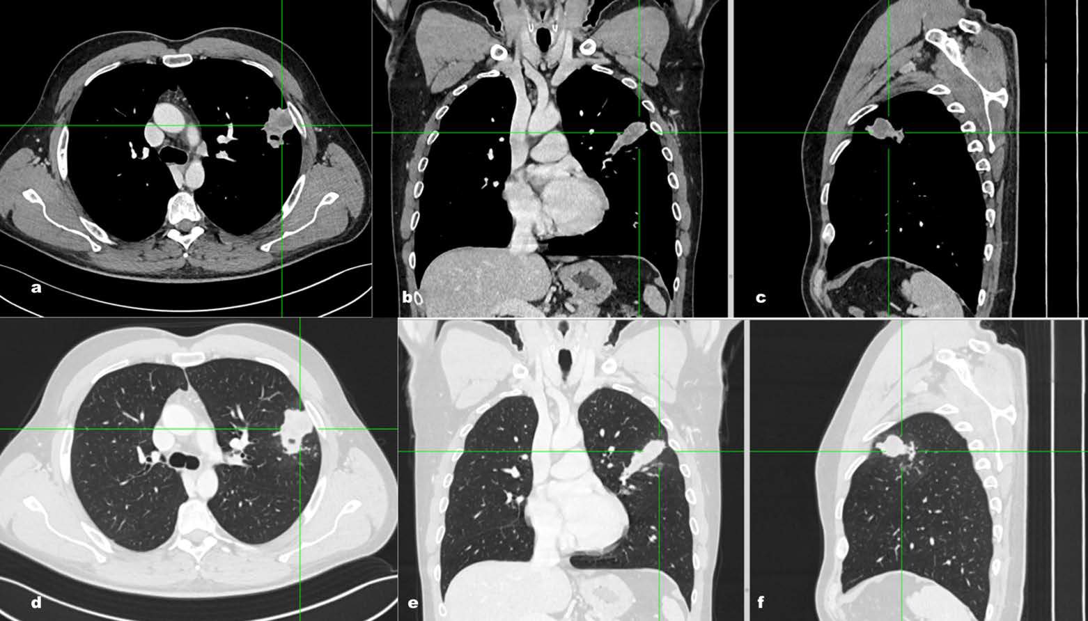
Eosinophilic pneumonia (EP) is a rare interstitial lung disease often mimicking other pulmonary conditions. We present the case of a 43-year-old male evaluated for suspected lung cancer due to progressive dyspnea, weight loss, cough. Cytological analysis confirmed an interstitial pulmonary inflammatory process with significant eosinophilic granulocyte infiltration, with no evidence of malignancy. Complete clinical and radiological resolution was achieved within two weeks following corticosteroid and empiric antibiotic therapy. This case highlights the challenging differential diagnosis of EP, especially when mimicking malignancy, emphasizing the crucial role of a complete clinical evaluation based on a multimodality imaging diagnostic assessment and prompt response to corticosteroids for diagnosis confirmation.
Downloads
Published
Issue
Section
License
Copyright (c) 2025 Journal of Radiology Case Reports

This work is licensed under a Creative Commons Attribution-NonCommercial-NoDerivatives 4.0 International License.
The publisher holds the copyright to the published articles and contents. However, the articles in this journal are open-access articles distributed under the terms of the Creative Commons Attribution-NonCommercial-NoDerivs 4.0 License, which permits reproduction and distribution, provided the original work is properly cited. The publisher and author have the right to use the text, images and other multimedia contents from the submitted work for further usage in affiliated programs. Commercial use and derivative works are not permitted, unless explicitly allowed by the publisher.


