
Osteoid Osteoma of the Joint Capsule: A Case Report Highlighting Diagnostic Challenges and the Role of Advanced Imaging
DOI:
https://doi.org/10.3941/jrcr.5804Abstract
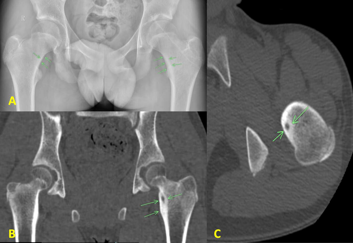
BACKGROUND: Osteoid osteoma is a benign osteogenic tumor typically affecting cortical bone and often mimicking musculoskeletal conditions. This report describes a rare intra-articular osteoid osteoma within the hip joint capsule, highlighting diagnostic challenges and the role of multimodal imaging.
A 13-year-old male presented with a one-year history of persistent proximal left femoral pain, most severe at night, disrupting sleep. Initial computed tomography revealed a lesion extending from the proximal femoral cortex into the medullary cavity and quadratus femoris muscle, while contrast-enhanced magnetic resonance imaging confirmed an intra-capsular nidus. Surgical resection identified woven bone, confirming the diagnosis. This case demonstrates that computed tomography and contrast-enhanced magnetic resonance imaging are essential for visualizing the nidus and reducing misdiagnosis in atypical cases.
Downloads
Published
Issue
Section
License
Copyright (c) 2025 Journal of Radiology Case Reports

This work is licensed under a Creative Commons Attribution-NonCommercial-NoDerivatives 4.0 International License.
The publisher holds the copyright to the published articles and contents. However, the articles in this journal are open-access articles distributed under the terms of the Creative Commons Attribution-NonCommercial-NoDerivs 4.0 License, which permits reproduction and distribution, provided the original work is properly cited. The publisher and author have the right to use the text, images and other multimedia contents from the submitted work for further usage in affiliated programs. Commercial use and derivative works are not permitted, unless explicitly allowed by the publisher.


