
Leiomyosarcoma of the Mediastinum Depicted on CT Images: A Rare Origin Case and Literature Review
DOI:
https://doi.org/10.3941/jrcr.5800Abstract
Introduction: Leiomyosarcoma (LMS), a malignant smooth muscle tumor, accounts for 10%–20% of soft tissue sarcomas. Primary mediastinal LMS is rare, with only 13 cases reported in the last two decades. This study aims to delineate its clinical and radiological characteristics through a novel case presentation and literature review. Methods: We report the case of a 57-year-old male presenting with progressive dyspnea and back pain. Diagnostic workup included contrastenhanced chest CT, histopathological analysis of CT-guided biopsy specimens, and immunohistochemical staining (positive for Vim, Des, SMA, and Calponin; negative for S100 and CD34). A systematic PubMed review of mediastinal LMS cases was performed to summarize imaging and pathological features.
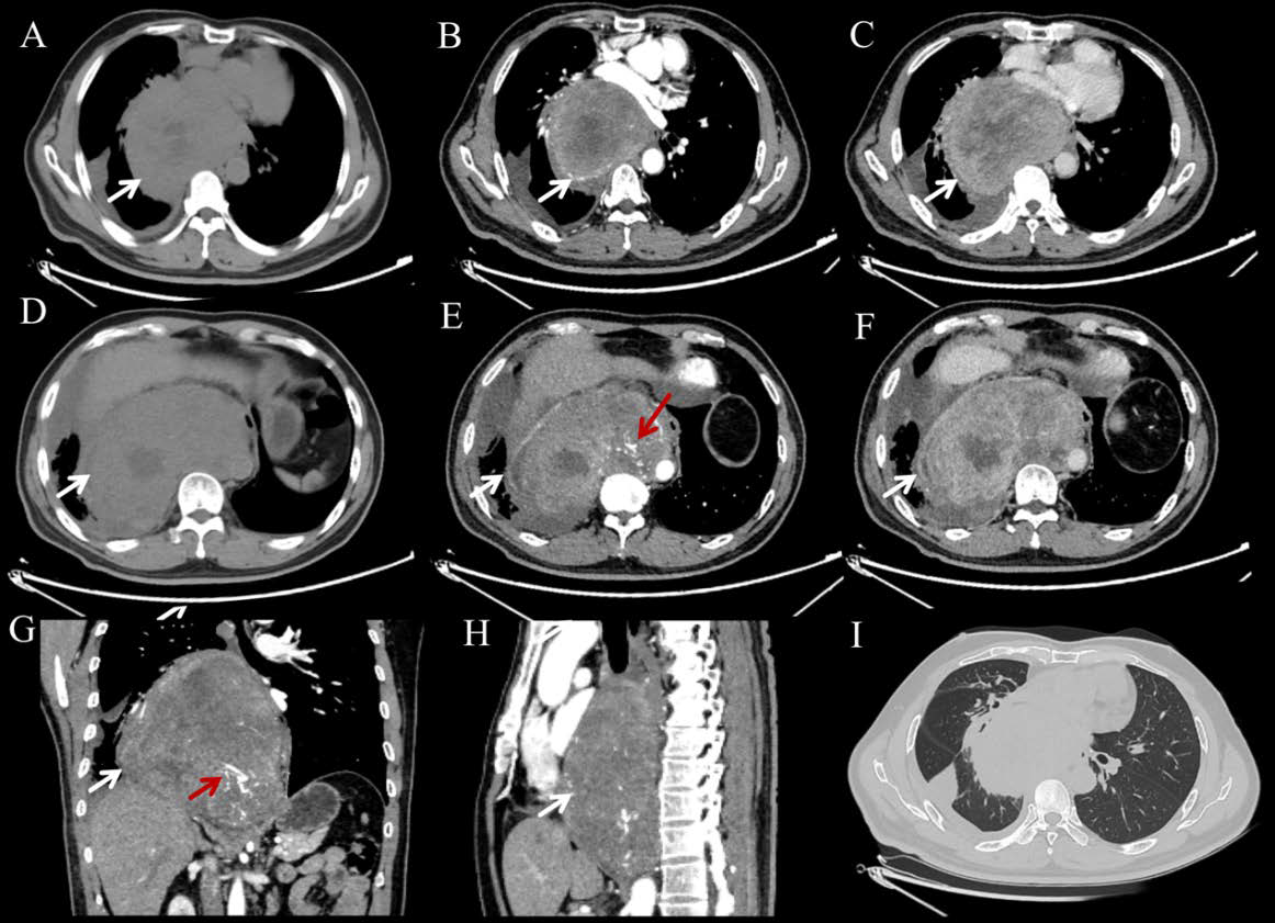
Results: Imaging revealed a 16.1 × 10.9 cm heterogeneous posterior mediastinal mass with necrotic foci, vascular encasement, and right pleural effusion. Histopathology showed spindle-shaped tumor cells with a Ki-67 index of 40%, confirming LMS. Literature synthesis (n=14 cases) demonstrated that 71.4% of tumors exceeded 5 cm, with a predilection for the anterior mediastinum (71.4%) and heterogeneous enhancement (100%).
Discussion: Mediastinal LMS radiologically mimics other malignancies but exhibits distinctive pathological markers (SMA/ Desmin+). Surgical resection remains the primary treatment; our patient received neoadjuvant chemotherapy (liposomal doxorubicin/dacarbazine) and achieved a partial response. This case highlights the importance of multidisciplinary collaboration (imaging-pathology correlation) for the timely diagnosis of this aggressive neoplasm.
Downloads
Published
Issue
Section
License
Copyright (c) 2025 Journal of Radiology Case Reports

This work is licensed under a Creative Commons Attribution-NonCommercial-NoDerivatives 4.0 International License.
The publisher holds the copyright to the published articles and contents. However, the articles in this journal are open-access articles distributed under the terms of the Creative Commons Attribution-NonCommercial-NoDerivs 4.0 License, which permits reproduction and distribution, provided the original work is properly cited. The publisher and author have the right to use the text, images and other multimedia contents from the submitted work for further usage in affiliated programs. Commercial use and derivative works are not permitted, unless explicitly allowed by the publisher.


