
False Positive Brown Adipose Tissue Uptake on Parathyroid 99mTc-Sestamibi Scan
DOI:
https://doi.org/10.3941/jrcr.5780Abstract
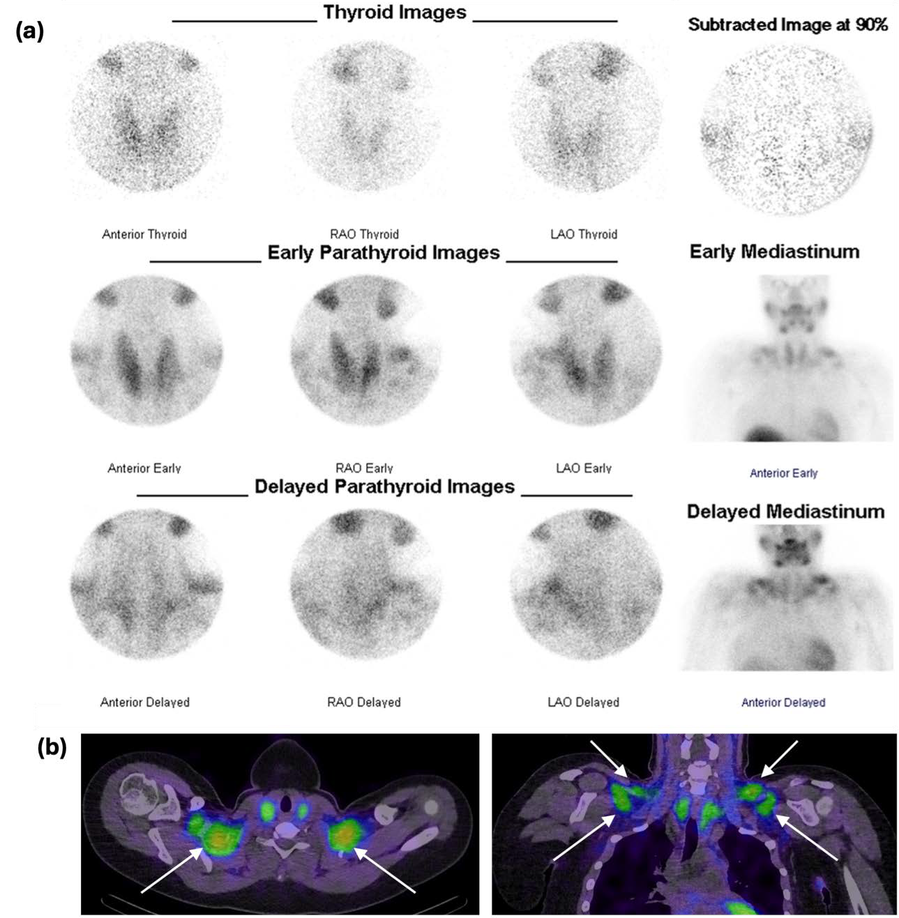
An 18-year-old female was found to have hyperparathyroidism and severe chronic renal failure of unclear cause. Dual-isotope parathyroid imaging was performed to exclude a parathyroid adenoma. Pinhole scintigraphy following intravenous administration of 99mTc-pertechnetate showed homogenous activity throughout the orthotopic thyroid gland. Early and delayed pinhole and planar scintigraphy following 99mTc-sestamibi administration showed discordant activity in the bilateral neck and supraclavicular regions, confirmed as brown adipose tissue on SPECT-CT. This case demonstrates false positive 99mTc-sestamibi uptake within brown adipose tissue in the neck and illustrates the value of SPECT-CT in differentiating brown adipose tissue from parathyroid activity.
Downloads
Published
Issue
Section
License
Copyright (c) 2025 Journal of Radiology Case Reports

This work is licensed under a Creative Commons Attribution-NonCommercial-NoDerivatives 4.0 International License.
The publisher holds the copyright to the published articles and contents. However, the articles in this journal are open-access articles distributed under the terms of the Creative Commons Attribution-NonCommercial-NoDerivs 4.0 License, which permits reproduction and distribution, provided the original work is properly cited. The publisher and author have the right to use the text, images and other multimedia contents from the submitted work for further usage in affiliated programs. Commercial use and derivative works are not permitted, unless explicitly allowed by the publisher.


