An Atypical Case of Splenic Lymphangioma
DOI:
https://doi.org/10.3941/jrcr.5672Abstract
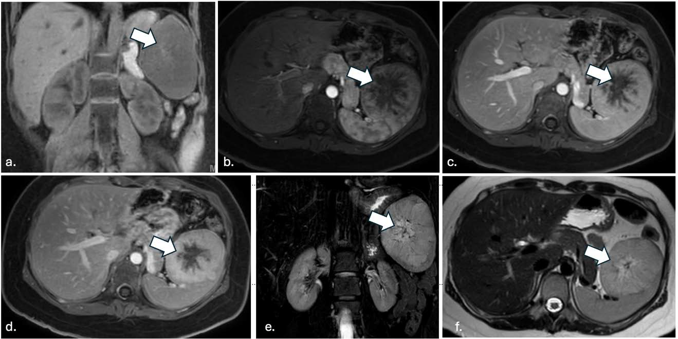
Lymphangioma is a rare, benign lymphatic malformation that most commonly occurs in infants and toddlers. It is often asymptomatic and detected incidentally on US or CT, though MRI allows optimal characterization. The spleen is an uncommon location for lymphangiomas, with the neck (75%) and axilla (20%) being the most common locations, followed by orbit, bone, mediastinum, and abdomen less commonly. When the lesions occur in the abdomen, they are most commonly found in the mesentery and omentum, in addition to the adrenal, kidney, gastrointestinal tract, liver, and pancreas. The lesions commonly are cystic with septa/loculations, and no hypermetabolism on PET. This report presents an atypical case of a young adult with a splenic lymphangioma, an already rare finding, which is further unusual in having a solid appearance with lack of septa/loculations, and mild hypermetabolism on PET, though it was confirmed as a lymphangioma with classic findings demonstrated on histopathology. There were also arm and back lesions that likely represented venous malformations based on radiology (no pathology was obtained), signifying a systemic process, and raised suspicion that the splenic lesion was of similar etiology. Genetic analysis identified a c.2740G>A mutation on exon 19 of the PIK3CA gene, in keeping with being on the PIK3CA-related overgrowth spectrum (PROS), which includes Klippel-Trenaunay syndrome, a rare disorder involving vascular and lymphatic malformations. This case highlights that splenic lymphangioma should be considered in cases with splenomegaly or left upper quadrant pain, and in the differential with hemangiomas and other splenic lesions, even in non-classic presentations clinically and on imaging. The presence of venous malformations elsewhere should also raise suspicion for splenic lesions being of similar nature and the possibility of a vascular malformation syndrome

Downloads
Published
Issue
Section
License
Copyright (c) 2025 Journal of Radiology Case Reports

This work is licensed under a Creative Commons Attribution-NonCommercial-NoDerivatives 4.0 International License.
The publisher holds the copyright to the published articles and contents. However, the articles in this journal are open-access articles distributed under the terms of the Creative Commons Attribution-NonCommercial-NoDerivs 4.0 License, which permits reproduction and distribution, provided the original work is properly cited. The publisher and author have the right to use the text, images and other multimedia contents from the submitted work for further usage in affiliated programs. Commercial use and derivative works are not permitted, unless explicitly allowed by the publisher.


