
Unilateral Internal Cerebral Vein Thrombosis: Imaging of an Uncommon Case and Literature Review
DOI:
https://doi.org/10.3941/jrcr.5636Abstract
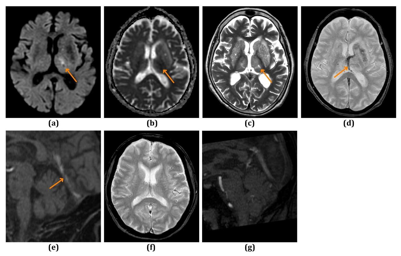
Cerebral venous thrombosis can lead to detrimental outcomes such as cerebral edema, hemorrhagic infarcts, neurological deficits, and death if not diagnosed and treated promptly. Patients with deep cerebral venous thrombosis face a poorer prognosis, with 29.4% experiencing lifelong dependence or death. Unilateral internal cerebral vein thrombosis is an uncommon and potentially life-threatening entity not widely published in literature. It can mimic arterial infarcts from artery of Percheron/basilar tip thrombosis, infectious, neoplastic and metabolic disorders. Radiologists must recognize its imaging features to ensure early diagnosis and timely treatment. In this article, we present the imaging findings of an uncommon case of unilateral internal cerebral vein thrombosis and how it can be distinguished from other diagnoses. We also review relevant literature, explore the tendency for left internal cerebral vein thrombosis, and discuss the diagnostic accuracy and pitfalls in imaging.
Downloads
Published
Issue
Section
License
Copyright (c) 2025 Journal of Radiology Case Reports

This work is licensed under a Creative Commons Attribution-NonCommercial-NoDerivatives 4.0 International License.
The publisher holds the copyright to the published articles and contents. However, the articles in this journal are open-access articles distributed under the terms of the Creative Commons Attribution-NonCommercial-NoDerivs 4.0 License, which permits reproduction and distribution, provided the original work is properly cited. The publisher and author have the right to use the text, images and other multimedia contents from the submitted work for further usage in affiliated programs. Commercial use and derivative works are not permitted, unless explicitly allowed by the publisher.


