
Intracranial Tumors Using Magnetic Resonance Spectroscopy
DOI:
https://doi.org/10.3941/jrcr.5578Abstract
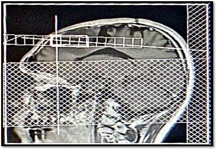
Magnetic resonance spectroscopy (MRS), which can identify and measure brain components (metabolites) in pathological lesions and normal-appearing tissues, offers a valuable additional diagnostic tool for assessing several neurological diseases. The study aimed to determine how MRS helps distinguish chemical shifts in brain lesions. Materials and Methods: Three MRS cases were retrospectively evaluated among patients from the Saudi German Hospital between February and April 2023. Results: In MRS, a high choline/creatinine ratio with a drop in NAA was indicative of a high T2 flare and illness progression, confirming a space-occupying lesion (SOL) after left frontal craniotomy. MRS curve analysis showed a small area of the reversed choline/N-acetyl aspartate (NAA/choline) curve. There is a perifocal area of altered signal that could present gliosis rather than edema due to the absence of significant positive mass defects. The final impression was right parieto-occipital diffuse astrocytoma (G2). The last case, MRS, has a low choline-to-creatinine ratio and is likely to be inactive in Baló concentric sclerosis. Conclusions: MRS is a crucial technique for analysis and routine monitoring of brain malignancies. MRS may be useful and helpful in some situations and may aid in determining the extent of the disease or the location of hotspots.
Downloads
Published
Issue
Section
License
Copyright (c) 2025 Journal of Radiology Case Reports

This work is licensed under a Creative Commons Attribution-NonCommercial-NoDerivatives 4.0 International License.
The publisher holds the copyright to the published articles and contents. However, the articles in this journal are open-access articles distributed under the terms of the Creative Commons Attribution-NonCommercial-NoDerivs 4.0 License, which permits reproduction and distribution, provided the original work is properly cited. The publisher and author have the right to use the text, images and other multimedia contents from the submitted work for further usage in affiliated programs. Commercial use and derivative works are not permitted, unless explicitly allowed by the publisher.


