
Case Report: Polysplenia Syndrome, a Wide Range of Congenital Heart Malformations in an Adult
DOI:
https://doi.org/10.3941/jrcr.5350Abstract
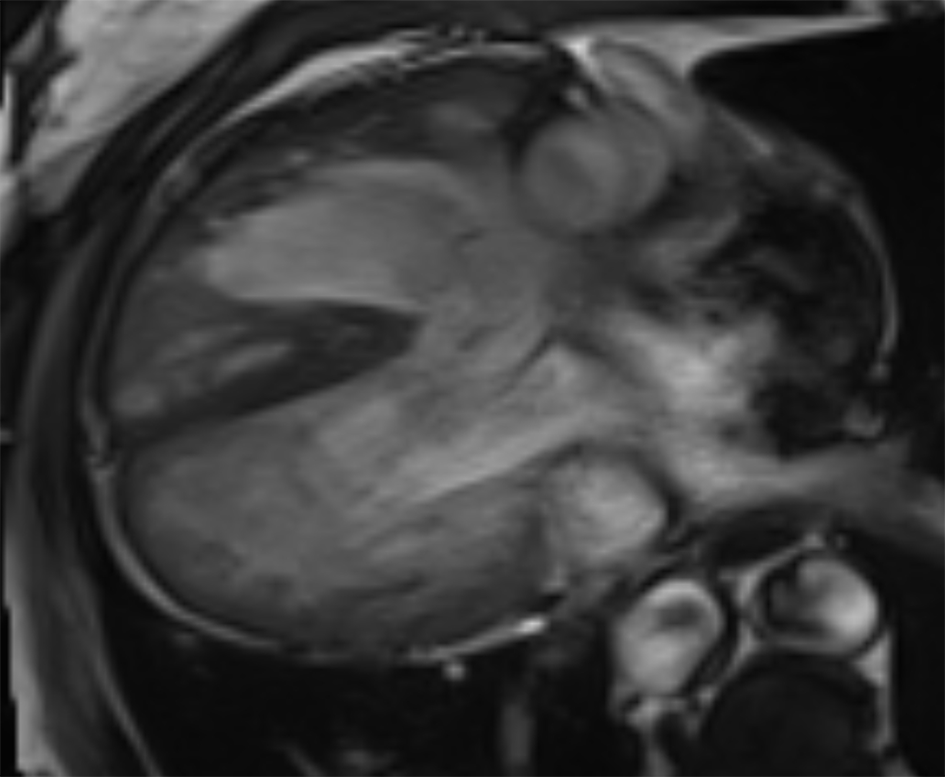
Background: Polysplenia syndrome is a rare disease where are observed two or more spleens associated with left-sided isomerism. In this condition there are many visceral and vascular congenital malformations, including cardiac anomalies.
Case summary: In this case presentation of a 60-year-old male with polysplenia syndrome, are identified multiple cardiovascular abnormalities and, despite these malformations, the patient survived with minor symptoms without surgery, highlighting the need for individualized management.
Discussion: Even if polysplenia syndrome is a rare condition, not easy to manage, this case report highlights the importance of a multidisciplinary medical board in the choice of the assessment, recognizing that some patient can have a good prognosis without surgical intervention.
Downloads
Published
Issue
Section
License
Copyright (c) 2025 Journal of Radiology Case Reports

This work is licensed under a Creative Commons Attribution-NonCommercial-NoDerivatives 4.0 International License.
The publisher holds the copyright to the published articles and contents. However, the articles in this journal are open-access articles distributed under the terms of the Creative Commons Attribution-NonCommercial-NoDerivs 4.0 License, which permits reproduction and distribution, provided the original work is properly cited. The publisher and author have the right to use the text, images and other multimedia contents from the submitted work for further usage in affiliated programs. Commercial use and derivative works are not permitted, unless explicitly allowed by the publisher.


