
The Value of 3D SPACE MRI in Differentiating between Sequestrated Lumbar Disc Herniation and Tumors: Two Cases and Literature
DOI:
https://doi.org/10.3941/jrcr.v18i1.5195Abstract
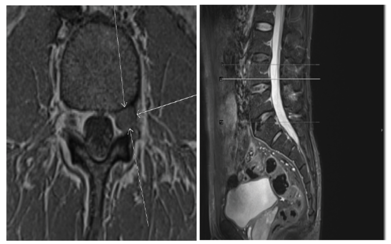
Background: Intervertebral disc herniation, defined as the protrusion or extrusion of the disc mass outside the disc space, is common and easy to diagnose on conventional Magnetic Resonance imaging (MRI) or Computed Tomography (CT) scans. However, the sequestrated disc fragments are challenging to detect, and intervertebral disc mass displacement into the dural sac, which can lead to serious neurological problems such as Cauda equina syndrome (CES). The sequestrated disc fragments do not have specific clinical or radiological characteristics that can differentiate an atypical disc mass from a tumor, making the diagnosis difficult preoperatively. Herein, we describe the use of Sampling Perfection with Application Optimized Contrast using different flip angle Evolution in Magnetic Resonance Imaging (3D SPACE MRI) in the diagnosis of the intervertebral disc fragment that mimicked a tumor.
Case presentation: In this study, we report two cases of sequestered lumbar disc herniation. The first case was a 37-year-old patient with a 2-year history of intermittent left lower limb pain that aggravates with exercise and is relieved at rest, while the second case was a 42-year-old patient with a history of 40 days of numbness and pain in the left lower limb.
Conclusion: 3D SPACE MRI is a beneficial diagnostic imaging tool for discriminating between disc mass that mimics a tumor and a tumor before surgery. If the disc fragment mimicking a tumor can be identified before the operation, open surgical treatment won't be necessary for all patients.
Downloads
Published
Issue
Section
License
Copyright (c) 2024 Journal of Radiology Case Reports

This work is licensed under a Creative Commons Attribution-NonCommercial-NoDerivatives 4.0 International License.
The publisher holds the copyright to the published articles and contents. However, the articles in this journal are open-access articles distributed under the terms of the Creative Commons Attribution-NonCommercial-NoDerivs 4.0 License, which permits reproduction and distribution, provided the original work is properly cited. The publisher and author have the right to use the text, images and other multimedia contents from the submitted work for further usage in affiliated programs. Commercial use and derivative works are not permitted, unless explicitly allowed by the publisher.


