
Blastomycosis of the Central Nervous System
DOI:
https://doi.org/10.3941/jrcr.v17i12.5167Abstract
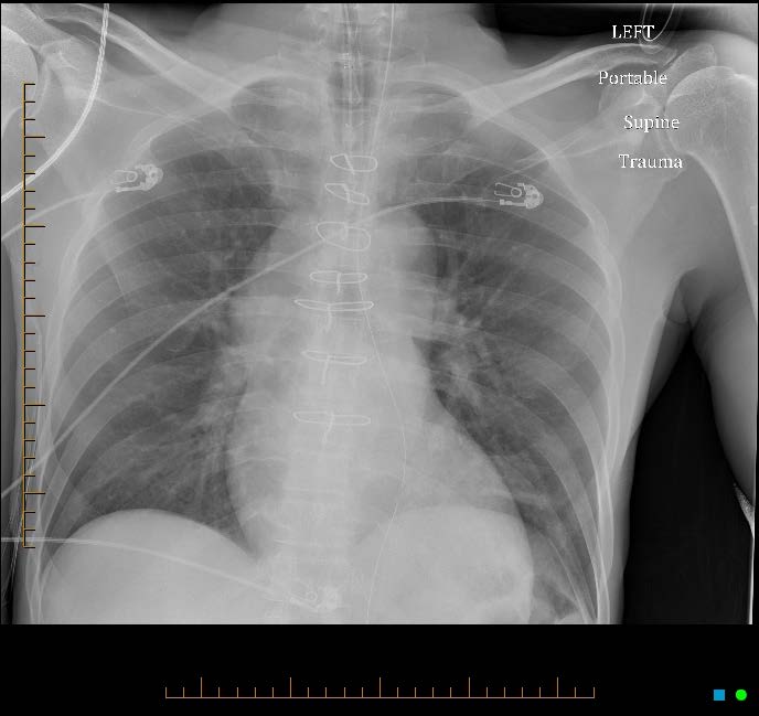
The reported incidence of blastomycosis is increasing in certain regions of the United States. The diagnosis is primarily made via urine antigen testing, culture, or cytology smear. The differential diagnosis for blastomycosis includes pneumonia, tuberculosis, and non-infectious pulmonary disease. Clinical context and epidemiologic exposure play a crucial role in diagnosis. However, the differential can expand significantly if there is disseminated central nervous system involvement, especially if pulmonary manifestations are not seen. Imaging begins to play a vital role when differentiating disseminated blastomycosis from other etiologies such as malignancy. Herein we present a case of a 58-year-old male who presented with seizures and right sided gaze preference found to have disseminated central nervous system blastomycosis. In this article, we will discuss symptoms and imaging findings of disseminated blastomycosis to help guide diagnosis and management.
Downloads
Published
Issue
Section
License
Copyright (c) 2024 Journal of Radiology Case Reports

This work is licensed under a Creative Commons Attribution-NonCommercial-NoDerivatives 4.0 International License.
The publisher holds the copyright to the published articles and contents. However, the articles in this journal are open-access articles distributed under the terms of the Creative Commons Attribution-NonCommercial-NoDerivs 4.0 License, which permits reproduction and distribution, provided the original work is properly cited. The publisher and author have the right to use the text, images and other multimedia contents from the submitted work for further usage in affiliated programs. Commercial use and derivative works are not permitted, unless explicitly allowed by the publisher.


