
Nasal obstruction in a 14 year old girl caused by a huge middle turbinate mucocele appearing radiologically as an inverted papilloma
DOI:
https://doi.org/10.3941/jrcr.v17i9.4774Keywords:
Concha Bullosa Mucocele, Concha Bullosa, Middle Turbinate, Paediatric Nasal MassAbstract
Introduction: We present a case of a fourteen year old girl who presented with a large intra-nasal mass to the ENT team at a district general hospital in the UK.
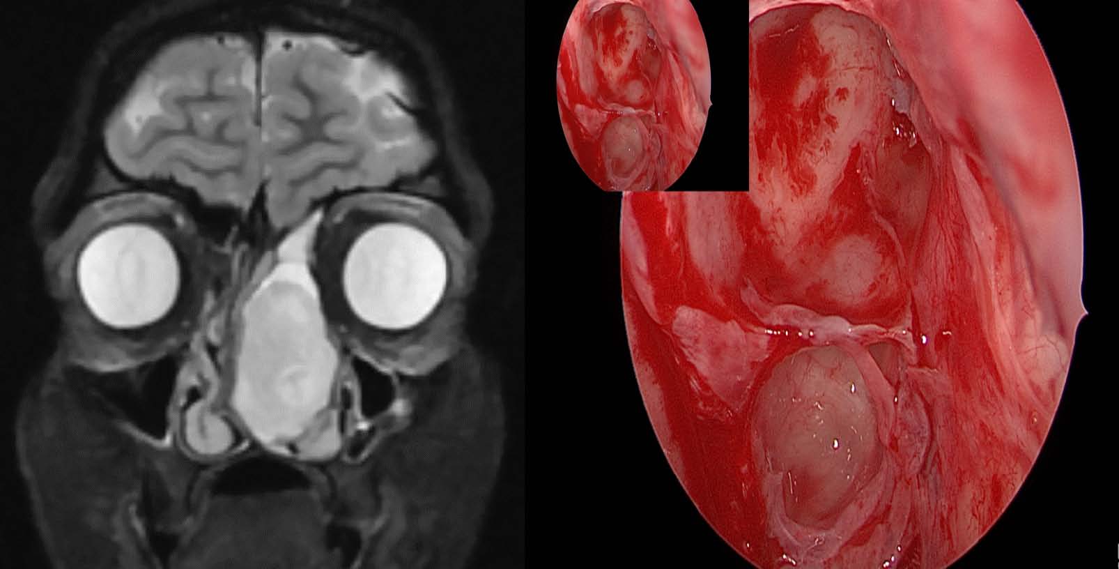
Presentation of case: The girl presented predominantly with nasal obstruction and some symptoms of allergic rhinitis. Imaging revealed a large lesion abutting the skull base and causing bony remodelling with marked septal deviation. Based both on CT and MRI imaging, the reporting (non-head and neck) radiologist suggested inverted papilloma as a differential diagnosis. Intra-operative exploration in fact revealed a very large left middle turbinate mucocele extending to the left frontal sinus. The mass was excised endoscopically without complications.
Discussion: Although concha bullosa of the middle turbinate of the nose are common, development of a mucocele within them is far less common and for such a mucocele to develop to this size in a child is extremely rare. The egg shell lining of the lesion can be a tell-tale sign of their aetiology when taken alongside other radiological factors. This case highlights challenges in radiological diagnosis of intra-nasal masses in children, which can lead to delays and increased anxiety.
Conclusion: When assessing nasal masses in children it is important to keep a wide differential due to the challenges of diagnosis. A close conversation should be had with local head and neck radiologists and, of course, where there is a unilateral nasal mass tissue sampling is essential and may be taken as part of a full excision where clinically indicated.
Downloads
Published
Issue
Section
License
Copyright (c) 2023 Journal of Radiology Case Reports

This work is licensed under a Creative Commons Attribution-NonCommercial-NoDerivatives 4.0 International License.
The publisher holds the copyright to the published articles and contents. However, the articles in this journal are open-access articles distributed under the terms of the Creative Commons Attribution-NonCommercial-NoDerivs 4.0 License, which permits reproduction and distribution, provided the original work is properly cited. The publisher and author have the right to use the text, images and other multimedia contents from the submitted work for further usage in affiliated programs. Commercial use and derivative works are not permitted, unless explicitly allowed by the publisher.


