
Sub-Lobar Dysplasia: Neuroimaging and Associated Histopathological Features
DOI:
https://doi.org/10.3941/jrcr.v18i2.4752Keywords:
Sublobar dysplasia, Cortical malformation, Neuroimaging, HistopathologyAbstract
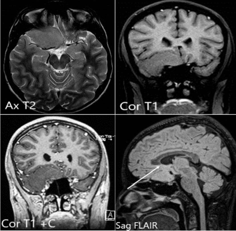
Sublobar dysplasia is a rare form of cortical malformation. It has been identified in the literature as having unique imaging findings and histopathological features. We report a rare case of sublobar dysplasia in the right frontal lobe, associated with detailed neuroimaging findings. Histopathological examination ruled out other types of dysplasia, underlying space-occupying lesions, and inflammatory processes.
Downloads
Published
Issue
Section
License
Copyright (c) 2024 Journal of Radiology Case Reports

This work is licensed under a Creative Commons Attribution-NonCommercial-NoDerivatives 4.0 International License.
The publisher holds the copyright to the published articles and contents. However, the articles in this journal are open-access articles distributed under the terms of the Creative Commons Attribution-NonCommercial-NoDerivs 4.0 License, which permits reproduction and distribution, provided the original work is properly cited. The publisher and author have the right to use the text, images and other multimedia contents from the submitted work for further usage in affiliated programs. Commercial use and derivative works are not permitted, unless explicitly allowed by the publisher.


