
Persistent craniopharyngeal canal with an associated sphenoid sinus fistula
DOI:
https://doi.org/10.3941/jrcr.v17i10.4707Keywords:
Craniopharyngeal Canal, Skull Base, CT, MRIAbstract
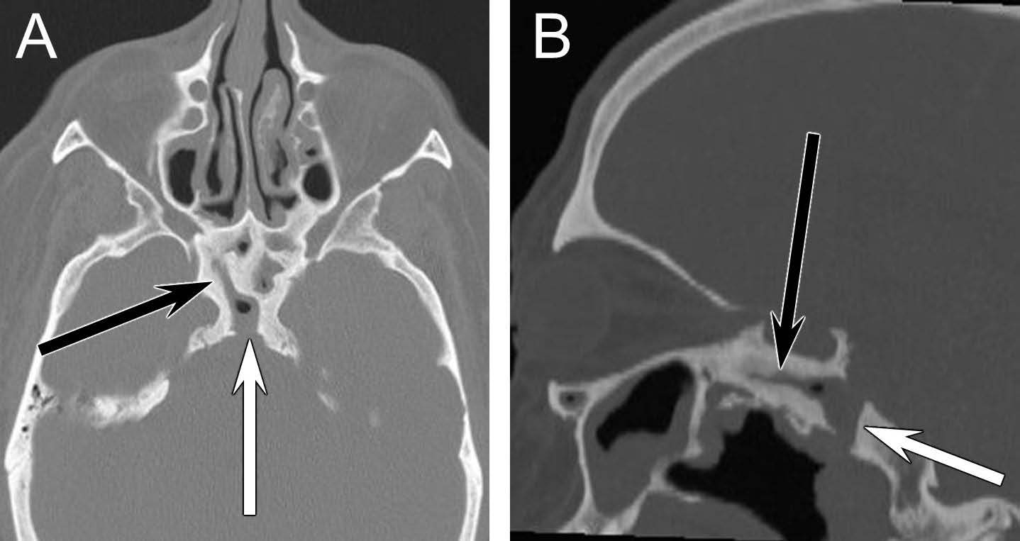
Persistent craniopharyngeal canal (PCC) is a rare congenital anomaly that appears as a linear well-corticated canal running from the sella through the clivus and into the nasopharynx. Case reports of this anomaly have shown it is associated with a range of craniofacial defects, pituitary abnormalities, and meningoencephaloceles. It predisposes patients to bacterial meningitis. In this case a 46-year-old gentleman presenting for preoperative planning for surgical drainage of Potts Puffy tumor was found to have a PCC on CT and MRI. Imaging also demonstrated the presence of chronic inflammation and a fistula extending from the tract into the sphenoid sinus. This unusual presentation of a PCC with a sphenoid sinus fistula broadens the potential clinical presentations of PCC and further emphasizes the ability of this anomaly to serve as a conduit for CNS infection.
Downloads
Published
Issue
Section
License
Copyright (c) 2023 Journal of Radiology Case Reports

This work is licensed under a Creative Commons Attribution-NonCommercial-NoDerivatives 4.0 International License.
The publisher holds the copyright to the published articles and contents. However, the articles in this journal are open-access articles distributed under the terms of the Creative Commons Attribution-NonCommercial-NoDerivs 4.0 License, which permits reproduction and distribution, provided the original work is properly cited. The publisher and author have the right to use the text, images and other multimedia contents from the submitted work for further usage in affiliated programs. Commercial use and derivative works are not permitted, unless explicitly allowed by the publisher.


