
Right Ventricular Involvement of an Aggressive Malignant Peripheral Nerve Sheath Tumor
DOI:
https://doi.org/10.3941/jrcr.v16i1.4338Keywords:
Malignant peripheral nerve sheath tumor, cardiac magnetic resonance imaging, transthoracic echocardiography, peripheral magnetic resonance imaging, cardiac metastasisAbstract
We present a case of a 58-year-old woman who had a painful right thigh mass for a few months. A transthoracic echocardiogram revealed no evidence of an intracardiac mass. She had a whole-body positron emission tomography/computed tomography scan two months later that revealed masses in her right lower extremity and a mass in her right ventricle that had not been initially reported. She had been initially diagnosed with an undifferentiated pleomorphic sarcoma, but this diagnosis was changed to a malignant peripheral nerve sheath tumor with repeat pathology. She was subsequently hospitalized. An echocardiogram showed a mass covering 80% of her right ventricle (RV). Serial cardiac magnetic resonance imaging revealed a 9.4 x 5.6 cm RV mass with vascular and avascular portions and inflow and outflow tract obstruction. Computed tomography showed no other metastases. Due to a delay in diagnosis and a decline in left ventricular ejection fraction, the patient could not undergo palliative chemotherapy or radiotherapy.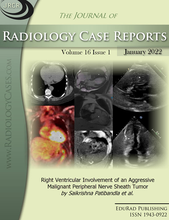
Downloads
Published
2022-01-31
Issue
Section
Cardiac Imaging
License
The publisher holds the copyright to the published articles and contents. However, the articles in this journal are open-access articles distributed under the terms of the Creative Commons Attribution-NonCommercial-NoDerivs 4.0 License, which permits reproduction and distribution, provided the original work is properly cited. The publisher and author have the right to use the text, images and other multimedia contents from the submitted work for further usage in affiliated programs. Commercial use and derivative works are not permitted, unless explicitly allowed by the publisher.


