
Long-Term Lumbar Multifidus Muscle Atrophy Changes Documented With Magnetic Resonance Imaging: A Case Series
DOI:
https://doi.org/10.3941/jrcr.v8i5.1401Keywords:
Lumbar multifidus muscle, lumbar multifidus muscle atrophy, multifidi, chronic low back pain, MRIAbstract
A retrospective case series of three patients with chronic low back pain who received baseline MRI scans revealing multifidus muscle atrophy with fatty replacement is provided. Each patient received spinal manipulative therapy, and two were compliant with low back exercises targeting the multifidus. A follow-up scan performed >1 year later was compared to the baseline scan revealing a decrease in atrophy with fatty replacement in the two patients who performed multifidus-focused low back exercises (15% and 39% on the left and 7% and 32% on the right respectively), and an increase in the patient who underwent spinal manipulation alone (41% and 53%). Interestingly, the decrease in atrophy in the two patients that performed the exercises correlated to functional improvements. Though limited, these results highlight the utility of MRI in quantifying positive and negative long-term changes in multifidus atrophy, which may be an indicator of recovery in chronic low back pain patients.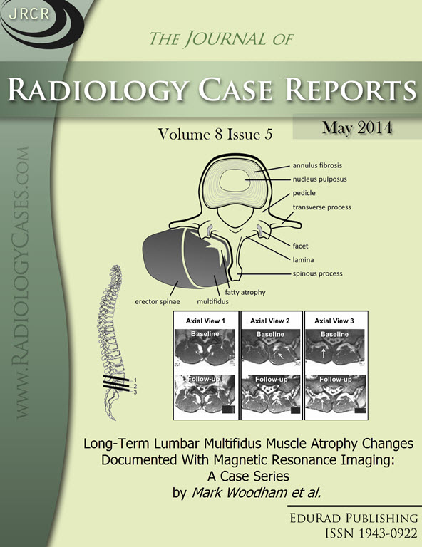
Downloads
Published
2014-05-25
Issue
Section
Musculoskeletal Radiology
License
The publisher holds the copyright to the published articles and contents. However, the articles in this journal are open-access articles distributed under the terms of the Creative Commons Attribution-NonCommercial-NoDerivs 4.0 License, which permits reproduction and distribution, provided the original work is properly cited. The publisher and author have the right to use the text, images and other multimedia contents from the submitted work for further usage in affiliated programs. Commercial use and derivative works are not permitted, unless explicitly allowed by the publisher.


