
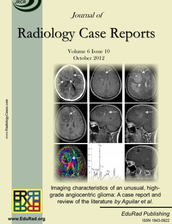
Imaging characteristics of an unusual, high-grade angiocentric glioma: A case report and review of the literature.
DOI:
https://doi.org/10.3941/jrcr.v6i10.1134Keywords:
angiocentric glioma, MR imaging, MR tractography, diffusion tensor imaging, MR spectroscopyAbstract
Angiocentric gliomas have recently been reclassified as a separate central nervous system tumor. Few cases have been reported, and most of those correspond to slow-growing, low-grade neoplasms in very young pediatric patients. Here we describe magnetic resonance imaging findings (including diffusion imaging, spectroscopy and tractography) in an unusual higher-grade neoplasm with pathologic features suggestive of an angiocentric glioma in a 15-year-old male. The tumor had mild heterogeneous enhancement on magnetic resonance imaging, and a low apparent diffusion coefficient (9.9 x 10⁻⁴ mm2s-1), consistent with an intermediate-to-high cellularity tumor. Spectroscopic imaging showed an elevated choline/phosphocreatine and choline/N-acetyl aspartate ratios, suggesting an unusually aggressive tumor. We conclude that angiocentric glioma should not be excluded from consideration at primary diagnosis, particularly in teenaged patients nearing adulthood.
Downloads
Published
Issue
Section
License
The publisher holds the copyright to the published articles and contents. However, the articles in this journal are open-access articles distributed under the terms of the Creative Commons Attribution-NonCommercial-NoDerivs 4.0 License, which permits reproduction and distribution, provided the original work is properly cited. The publisher and author have the right to use the text, images and other multimedia contents from the submitted work for further usage in affiliated programs. Commercial use and derivative works are not permitted, unless explicitly allowed by the publisher.


