
Superficial peroneal nerve compression due to peroneus brevis muscle herniation
DOI:
https://doi.org/10.3941/jrcr.v13i11.3757Keywords:
Peripheral Neuropathy, Muscle Hernia, Magnetic Resonance Imaging, Fasciotomy, Nerve ReleasingAbstract
Muscle hernias of the extremities most commonly occur in the leg, between the knee and ankle. Symptomatic muscle hernias in the leg are rare cause of chronic leg pain and neuropathy, and not routinely encountered in surgical practice. Although this condition is especially an esthetic problem, with palpable subcutaneous soft tissue mass, it can lead to spontaneous pain, cramp, local tenderness or potentially neuropathic symptoms. Moreover, among leg muscles involved in this process, peroneus brevis is less frequent than tibialis anterior. Magnetic Resonance Imaging is the method of choice in establishing the diagnosis. Symptomatic cases can be treated surgically in different ways, the preferred one is nerve releasing with fasciotomy. The purpose of this case report is to present the Magnetic Resonance findings of a superficial nerve compression due to a peroneus brevis muscle herniation.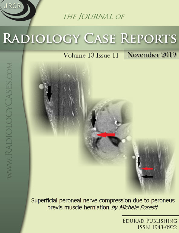
Downloads
Published
2019-11-22
Issue
Section
Musculoskeletal Radiology
License
The publisher holds the copyright to the published articles and contents. However, the articles in this journal are open-access articles distributed under the terms of the Creative Commons Attribution-NonCommercial-NoDerivs 4.0 License, which permits reproduction and distribution, provided the original work is properly cited. The publisher and author have the right to use the text, images and other multimedia contents from the submitted work for further usage in affiliated programs. Commercial use and derivative works are not permitted, unless explicitly allowed by the publisher.


