
Unusual Aggressive Breast Cancer: Metastatic Malignant Phyllodes Tumor
DOI:
https://doi.org/10.3941/jrcr.v7i2.1430Keywords:
phyllodes, cystosarcoma phyllodes, breast cancer, mesenchyal breast tumor, metastatic, enhancing septationsAbstract
For the year of 2012, it has been estimated that breast cancer will account for the greatest number of newly diagnosed cancers and the second highest proportion of cancer related deaths among women. Breast cancer, while often lumped together as one disease, represents a diverse group of malignancies with different imaging findings, histological appearances and behavior. While most invasive primary breast cancers are epithelial derived adenocarcinomas, rare neoplasms such as the phyllodes tumor may arise from mesenchymal tissue. Compared to the breast adenocarcinoma, the phyllodes tumor tends to affect a younger population, follows a different clinical course, is associated with different imaging and histological findings and is managed distinctively. There may be difficulty in differentiating the phyllodes tumor from a large fibroadenoma, but the mammographer plays a key role in reviewing the clinical and imaging data in order to arrive at the correct diagnosis. Early diagnosis with proper surgical management can often cure non-metastatic phyllodes tumors. However, in rare cases where metastasis occurs, prognosis tends to be poor. This report describes the presentation, imaging findings and management of a metastatic malignant phyllodes tumor.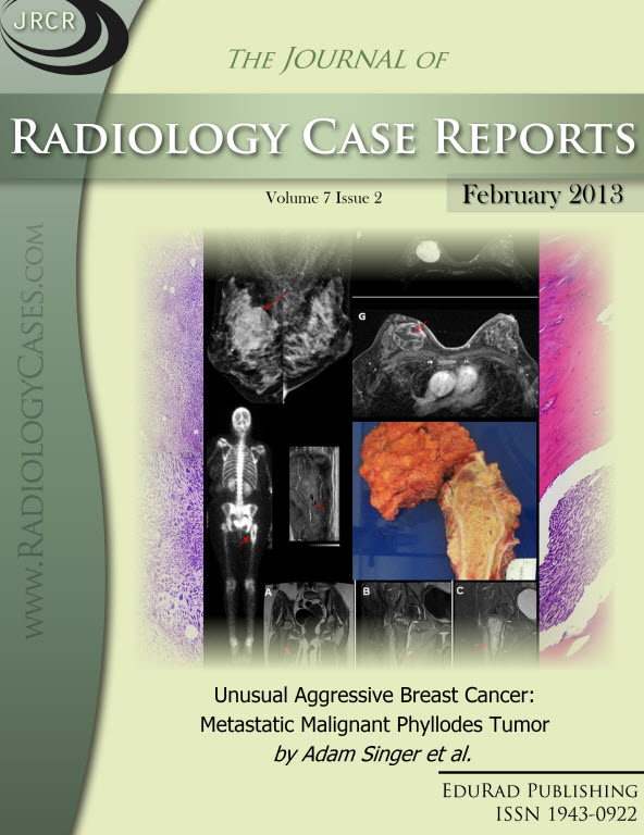
Downloads
Published
2013-02-17
Issue
Section
Breast Imaging
License
The publisher holds the copyright to the published articles and contents. However, the articles in this journal are open-access articles distributed under the terms of the Creative Commons Attribution-NonCommercial-NoDerivs 4.0 License, which permits reproduction and distribution, provided the original work is properly cited. The publisher and author have the right to use the text, images and other multimedia contents from the submitted work for further usage in affiliated programs. Commercial use and derivative works are not permitted, unless explicitly allowed by the publisher.


