
Involvement of the Mediastinal Subpleural Pulmonary Parenchyma on Chest CT in COVID-19 patients: A Case Series
DOI:
https://doi.org/10.3941/jrcr.v14i11.3974Keywords:
Coronavirus disease 2019, COVID-19, chest CT, ground-glass opacities, GGOs, lung consolidation, mediastinal pleuraAbstract
Coronavirus disease 2019 (COVID-19) is an infectious disease caused by the severe acute respiratory syndrome coronavirus 2 (SARS-CoV-2). First identified in December 2019 in Wuhan, China, it has since become a global pandemic. Although the reference standard for SARS-CoV-2 diagnosis is real-time reverse transcription polymerase chain reaction (RT-PCR), computed tomography (CT) is recommended for both initial evaluation and follow-up. The CT findings in COVID-19 are varied, but typical ground-glass opacities are usually reported to occupy a peripheral costal subpleural distribution. Here we report eight confirmed COVID-19 cases who underwent clinical evaluation, laboratory testing, and unenhanced chest CT. In all patients, chest CT showed the presence of ground-glass opacities in the mediastinal subpleural parenchyma. While these cases also showed the typical CT features of COVID-19, involvement of the mediastinal subpleural parenchyma should not lower the index of suspicion for COVID-19.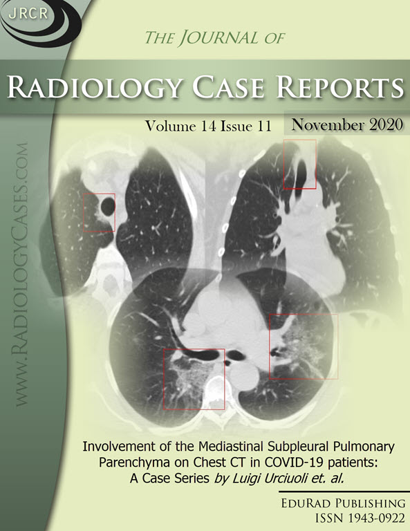
Downloads
Published
2020-11-28
Issue
Section
Thoracic Radiology
License
The publisher holds the copyright to the published articles and contents. However, the articles in this journal are open-access articles distributed under the terms of the Creative Commons Attribution-NonCommercial-NoDerivs 4.0 License, which permits reproduction and distribution, provided the original work is properly cited. The publisher and author have the right to use the text, images and other multimedia contents from the submitted work for further usage in affiliated programs. Commercial use and derivative works are not permitted, unless explicitly allowed by the publisher.


