
Radiological Findings in Persistent Mí¼llerian Duct Syndrome: Case Report and Review of Literature
DOI:
https://doi.org/10.3941/jrcr.v11i3.3027Keywords:
PMDS, Persistent Mí¼llerian duct syndrome, Hernia uteri inguinalis, Mí¼llerian duct, Wolffian duct, Scrotal ultrasound, Abdominal CT, Abdominal MRIAbstract
This case involved a 36-year-old adult male who presented with an unusual inguinal hernia in which the uterus and fallopian tubes were identified as contents of the inguinal hernia sac. These findings reflected a rare autosomal recessive developmental syndrome known as PMDS (persistent Mí¼llerian duct syndrome). The diagnosis was established and confirmed via radiological-mainly MRI-investigation.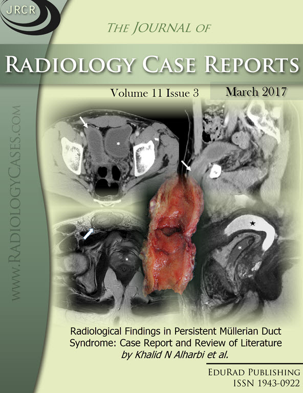
Downloads
Published
2017-03-23
Issue
Section
Genitourinary Radiology
License
The publisher holds the copyright to the published articles and contents. However, the articles in this journal are open-access articles distributed under the terms of the Creative Commons Attribution-NonCommercial-NoDerivs 4.0 License, which permits reproduction and distribution, provided the original work is properly cited. The publisher and author have the right to use the text, images and other multimedia contents from the submitted work for further usage in affiliated programs. Commercial use and derivative works are not permitted, unless explicitly allowed by the publisher.


