
Incisional Hernia Following Ventriculoperitoneal Shunt Positioning
DOI:
https://doi.org/10.3941/jrcr.v10i6.2329Keywords:
Ventriculoperitoneal shunt, incisional hernia, shunt dislocation, Ultrasonography, US, Sonography, CT, X-ray, abdomen, hydrocephalusAbstract
Incisional hernia represents a rare complication after ventriculoperitoneal shunt positioning due to failure of the fascial suture in the site of abdominal entrance of ventriculoperitoneal catheter. Clinical presentation can be extremely variable, according to patient's performance status, herniated material constitution (i.e. mesenteric fat, bowel loops or both) and complication occurrence (e.g. strangulation or intestinal obstruction). Early diagnosis is fundamental in order to surgically repair the defect and prevent further complications. We present the case of a paucisymptomatic incisional hernia following ventriculoperitoneal shunt positioning. Diagnosis was made by means of ultrasound and confirmed by means of computed tomography. The patient was successfully managed by means of surgical repositioning of herniated loop and re-suture.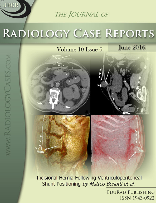
Downloads
Published
2016-06-24
Issue
Section
Gastrointestinal Radiology
License
The publisher holds the copyright to the published articles and contents. However, the articles in this journal are open-access articles distributed under the terms of the Creative Commons Attribution-NonCommercial-NoDerivs 4.0 License, which permits reproduction and distribution, provided the original work is properly cited. The publisher and author have the right to use the text, images and other multimedia contents from the submitted work for further usage in affiliated programs. Commercial use and derivative works are not permitted, unless explicitly allowed by the publisher.


